Computed tomography is commonly referred to as a CT scan. A CT scan is a diagnostic imaging procedure that uses a combination of X-rays and computer technology to produce images of the inside of the body. 
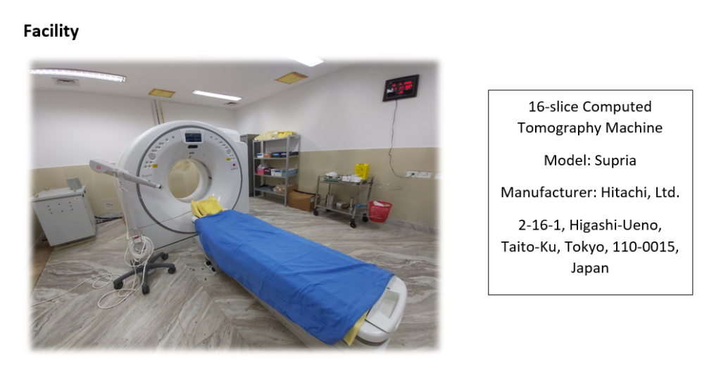
It shows detailed images of any part of the body, including the bones, muscles, fat, organs and blood vessels.

Background

The Computed tomography (CT) machine in the ERRH, Mongar is Hitachi Supria 16 slice. It was donated by JICA and was installed in August 2018.  The CT unit offers routine imaging services from 9 am to 3 pm on weekdays and 9 am to 1 pm on Saturday in addition 24×7 emergency services.

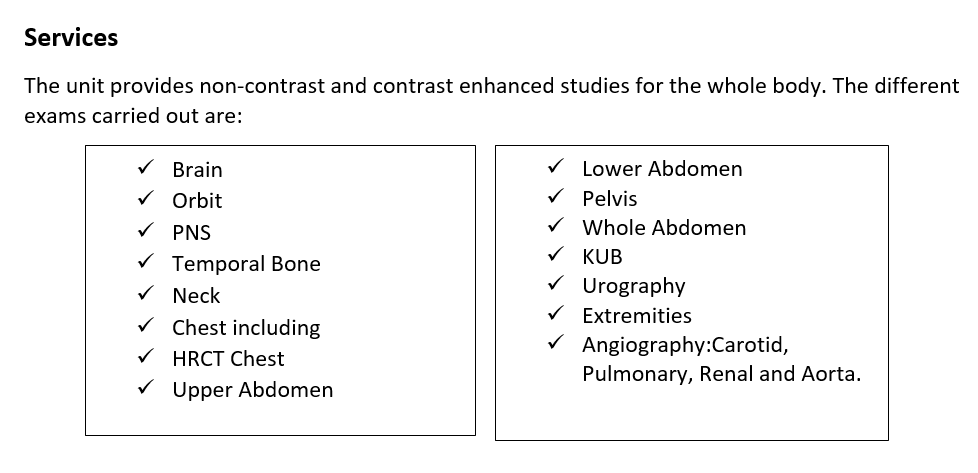
The CT unit gives all appointments along with the instruction on preparation for optimal imaging. The unit provides non-contrast and contrast enhanced studies for the whole body.

The CT unit headed by radiologist, who is also the head of the department, with two radio-technologists, two CT technicians and a CT nurse.

Facility

16-slice Computed Tomography Machine

Model: Supria

Manufacturer: Hitachi, Ltd. 2-16-1, Higashi-Ueno, Taito-Ku, Tokyo, 110-0015, Japan

Services

The unit provides non-contrast and contrast enhanced studies for the whole body. The different exams carried out are:

  • Brain
  • Orbit
  • PNS
  • Temporal Bone
  • Neck
  • Chest including HRCT Chest
  • Upper Abdomen
  • Lower Abdomen
  • Pelvis
  • Whole Abdomen
  • KUB
  • Urography
  • Extremities
  • Angiography: Carotid, Pulmonary, Renal and Aorta.

Faculty

  1. Dr. Kinley Sangay Dorji, Radiologist
  2. Mr. Karma Tshering, Radio-technologist
  3. Mr. Ugyen Phuntsho, Radio-technologist
  4. Mr. Kinzang Wangdi, CT Technician
  5. Mr. Rinzin Chophel, CT Technician
  6. Mr. Sherab Norbu, CT Nurse